
A corrective surgery performed in cases where the inner labia cause complaints due to size or asymmetry.
Genital Aesthetics About 45 minutes
Vaginoplasty (Vaginal Tightening)
Surgical repair of the vaginal canal and supporting tissues in cases of birth-related and age-related prolapse.
About Vaginoplasty (Vaginal Tightening)
Childbirth, age, and changes in tissue structure can lead to a sensation of widening in the vaginal canal. This condition can affect the quality of sexual life and self-confidence in some women. Vaginoplasty aims to surgically repair the loosened tissues and supporting structures; it is planned together with perineal repair if necessary. During the examination, the presence of accompanying conditions such as prolapse or urinary incontinence is evaluated, as these can alter the surgical plan. After surgery, sexual abstinence and certain restrictions are required throughout the recovery period. Results vary from person to person; no guaranteed success is promised, and expectations are discussed in detail before the operation.
Vaginoplasty is not a single standard surgery. The true anatomical cause of the complaint is identified; the surgery is personalized according to the level of support loss. You can click on the headings below to expand each section.
This page has been prepared taking into account the opinions of the American College of Obstetricians and Gynecologists (ACOG), guidelines from the National Institute for Health and Care Excellence (NICE) in the UK, studies published in PubMed Central and JAMA Network, as well as the 2026 Turkish national expert consensus.
Note: This information is prepared for general patient education purposes. Vaginal laxity, cystocele, rectocele, urinary incontinence, and defecation problems may require different treatments. Which procedure is appropriate can only be determined after a detailed gynecological and pelvic floor examination.
Common Questions
Frequently asked questions
Answers to the most common questions about Vaginoplasty (Vaginal Tightening).
The vagina is a highly flexible fibromuscular organ that works together with its mucosa, smooth muscle layer, connective tissue, and surrounding pelvic floor support structures. Vaginal deliveries, pregnancy, traumas to the pelvic floor tissues, connective tissue characteristics, aging, menopause, and certain pelvic floor problems can cause a sensation of looseness and widening in the vaginal canal or vaginal opening.
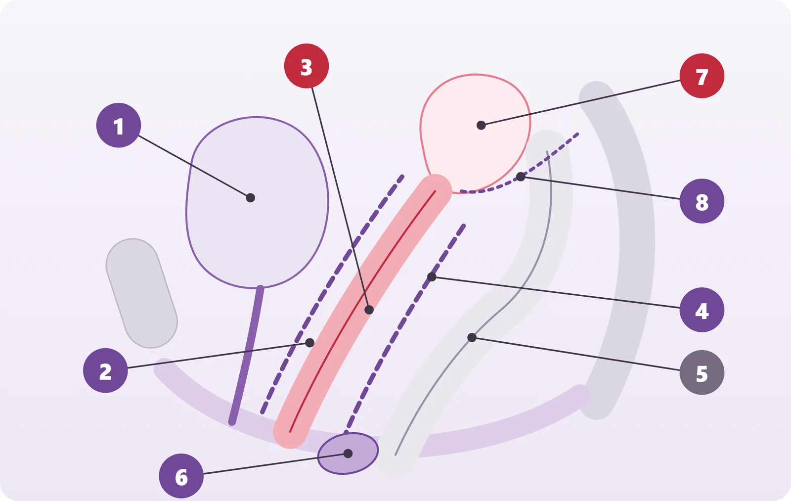
- Bladder
- Anterior wall (pubocervical) support tissues
- Vaginal canal
- Rectovaginal support (fascia–septum complex)
- Rectum
- Perineal body
- Uterus
- Apical (upper) support structures
In international urogynecology terminology, "vaginal laxity" is a symptom defined as a woman feeling her vagina is looser or wider than normal. This condition is reported more frequently especially in women who have had vaginal deliveries and may be associated with the widening of the levator hiatus, excessive distension of the pelvic floor muscles, and pelvic organ prolapse.
The term vaginoplasty has a broad meaning in medicine. When used in the context of genital aesthetics and pelvic reconstructive surgery, it generally refers to reducing the width of the vaginal canal and rearranging the support structures. "Vaginal rejuvenation" is not a standard medical diagnosis but a broader term that can be used for marketing purposes. ACOG notes that some vaginal tightening procedures can significantly overlap with classic anterior or posterior colporrhaphy techniques.
There is an important distinction here:
The sensation of vaginal looseness is not the same as cystocele, rectocele, vaginal opening widening, or pelvic organ prolapse.
Therefore, in our clinic, "vaginoplasty" is not considered a single standard surgery. First, it is evaluated whether the looseness is at the vaginal opening or in the middle or upper vaginal canal; anterior wall bladder support, posterior wall rectum support, perineal muscles, and apical support are assessed separately.
Why are the vaginal support tissues important?
The connective tissue complex that provides support between the bladder and vagina on the anterior vaginal wall is generally associated with the pubocervical/endopelvic support tissues. On the posterior side, the rectovaginal connective tissue/fascia-septum complex lies between the vagina and rectum.
Weakening or defects in these structures can contribute to pelvic floor problems such as cystocele on the anterior wall and rectocele on the posterior wall.
Therefore, especially in patients with accompanying pelvic support disorders, the goal of successful surgery is not only to remove excess mucosa on the vaginal surface but to identify the affected support layer and perform anatomical reconstruction.
Vaginoplasty can be considered in women who experience vaginal looseness or a feeling of increased vaginal width that significantly affects their daily or sexual life, and in whom anatomical findings explaining this complaint are detected during examination.
Patients most commonly present with the following complaints:
- a noticeable feeling of vaginal looseness,
- less contact or friction during intercourse,
- reduced vaginal grip sensation during sexual intercourse,
- sound caused by air escaping from the vagina,
- opening at the vaginal entrance,
- damage in the perineal area after childbirth or accompanying pelvic floor complaints.
The escape of air from the vagina is referred to in the literature as "vaginal flatus," "vaginal wind," or "garrulitas vulvae." It has been shown to be associated with vaginal delivery, a wide genital hiatus, levator ani muscle injury, and some pelvic organ prolapses.
In some women, the sensation of vaginal looseness may be accompanied by urinary incontinence, incomplete bladder emptying, pressure or fullness in the vaginal area, constipation, difficulty with bowel movements, or problems such as rectocele.
However, "vaginal tightening" alone should not be considered here. These complaints may be signs of cystocele, rectocele, stress urinary incontinence, or another pelvic floor disorder. Therefore, a woman requesting vaginoplasty in our clinic is evaluated not only for vaginal width but also in terms of urogynecological and pelvic floor anatomy.
Sexual satisfaction is not solely dependent on the diameter of the vagina. Clitoral stimulation, pelvic floor function, hormonal status, pain, lubrication, psychological, and relational factors also play roles in sexual function. Therefore, it is not scientifically accurate to guarantee that vaginoplasty will definitively increase sexual pleasure for every woman or her partner.
One of the strongest factors associated with vaginal laxity is pregnancy, especially vaginal delivery.
During childbirth, the vagina and pelvic floor tissues undergo significant distension. In some women, there may be stretching or avulsion of the levator ani muscles, widening of the genital hiatus, perineal injury, or weakening of the vaginal support tissues.
Recent studies have shown that women who have had vaginal deliveries report vaginal laxity complaints about three times more often compared to women who have delivered by cesarean section.
Other factors that may contribute to the weakening of vaginal and pelvic support structures over time include:
- aging,
- menopause and hormonal changes,
- connective tissue characteristics,
- obesity,
- chronic constipation and persistent straining,
- chronic cough,
- repeated increased intra-abdominal pressure and heavy physical exertion.
Rapid or large weight changes can alter the appearance of the external genital tissues and the volume of fat tissue. However, rapid weight loss alone cannot be said to be a proven primary cause of vaginal canal laxity. Therefore, anatomical causes should be determined by examination in each patient.
There is no single standard surgical technique applicable to all patients for vaginoplasty. The surgical method should be determined based on the location and degree of widening and any accompanying pelvic floor defects.
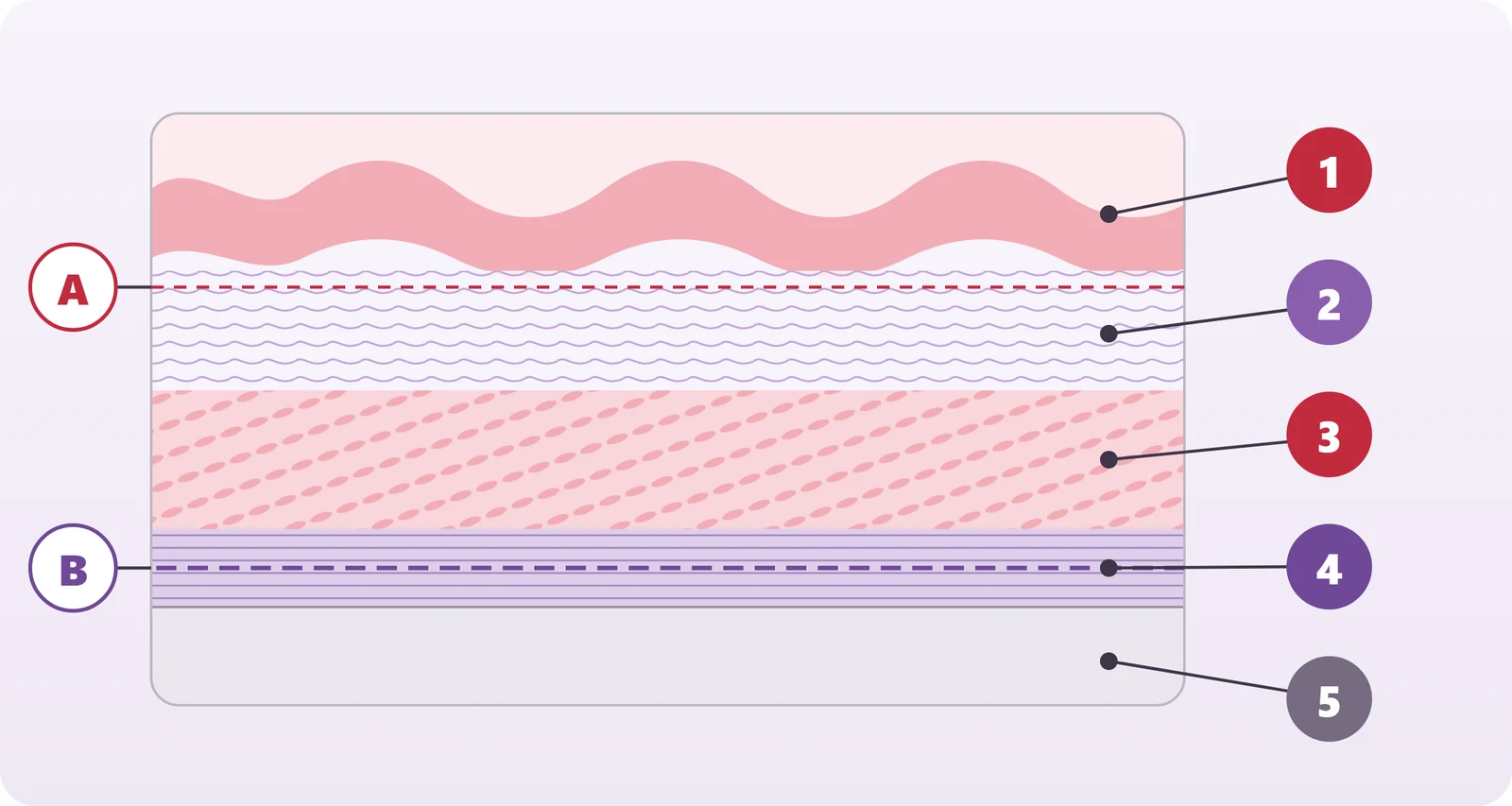
- Epithelium (non-keratinized stratified squamous epithelium)
- Lamina propria (collagen and elastic fibers)
- Smooth muscle layer
- Fascia and supportive tissue
- Adjacent organ (bladder on the anterior wall, rectum on the posterior wall)
- A — Superficial plane where only excess mucosa is removed
- B — Deep plane where supportive tissues are repaired
One of the most important principles in our clinic is:
Vaginal widening is not considered merely as the removal of excess vaginal mucosa.
The vaginal mucosa is histologically composed of non-keratinized stratified squamous epithelium. Beneath this lies the lamina propria rich in collagen and elastic fibers, smooth muscle layers, and connective tissue support. The vagina's capacity to expand during pregnancy, sexual intercourse, and childbirth arises not only from the epithelium but from this entire fibromuscular structure.
Therefore, removing only the mucosa may not correct the underlying true support defect.
In pelvic reconstructive surgery, the goal in anterior wall repairs is to evaluate and, if necessary, reconstruct the anterior supportive tissues; in posterior wall repairs, it is to assess and reconstruct the rectovaginal support complex. Excess mucosa can be removed in a controlled manner, but the structural part of the surgery involves repairing the underlying supportive tissues.
In our clinic, surgical planning is individualized for each patient. The areas of laxity along the vaginal canal, the regions with fascial support loss, cystocele, rectocele, or perineal defects are identified separately.
The approach we define as "total canal vaginoplasty" in our clinic is a surgical method based on evaluating the entire vaginal canal when necessary, rather than just narrowing a few centimeters of the vaginal entrance.
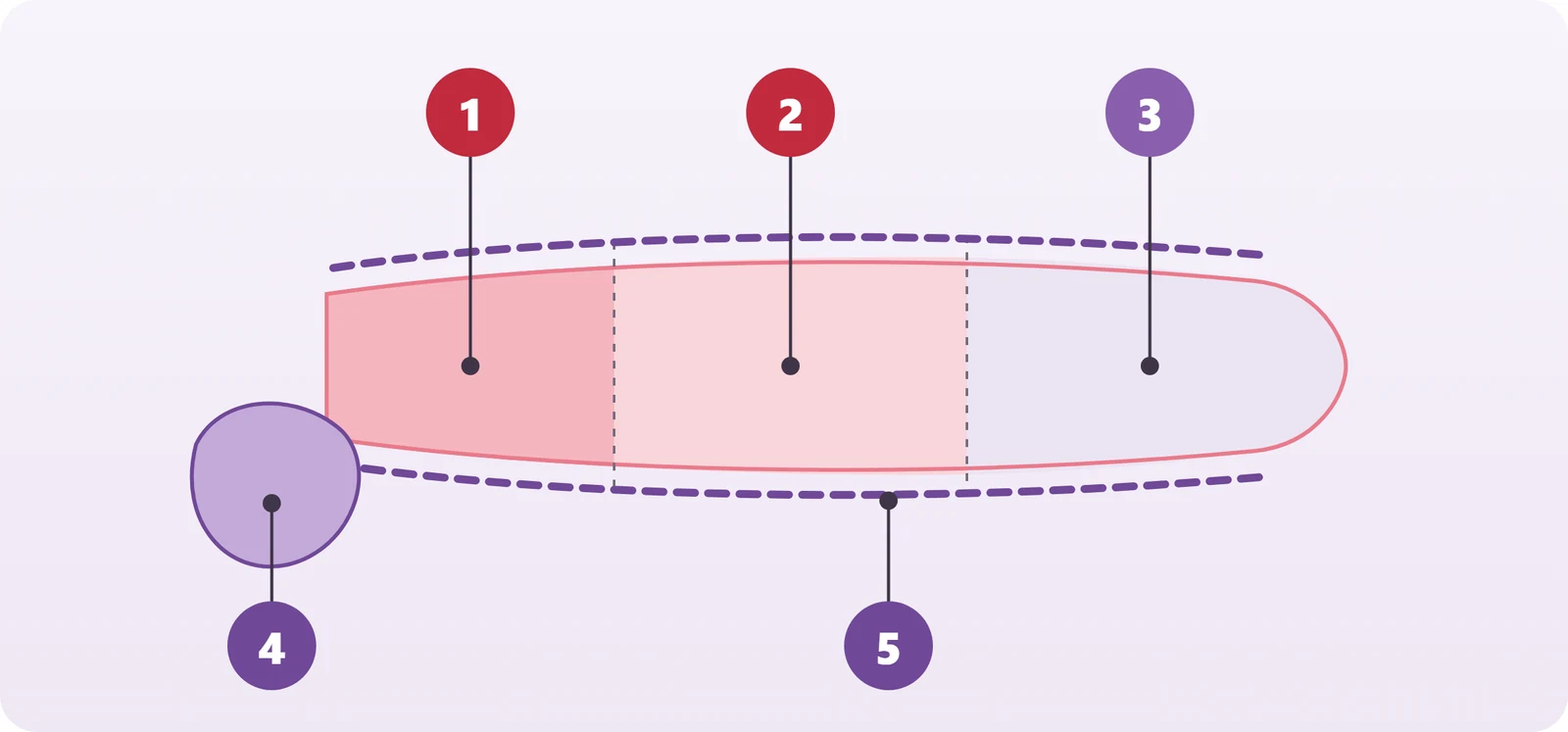
- Entrance (distal) level
- Middle level
- Upper (proximal) level
- Perineal body and entrance muscles
- Anterior and posterior wall support lines
The term "total canal" here does not mean the same procedure will be applied to every patient up to the cervix. The goal is to determine at which levels there is support loss from the vaginal entrance to the upper vaginal area and to plan the surgery according to the actual anatomical defect levels.
For example, if there is widening only at the vaginal entrance, a more limited perineal or distal repair may be sufficient. However, if there is support loss in the middle or proximal vaginal canal as well, narrowing only the vaginal entrance may not fully correct the problem.
In the national consensus study published in 2026 originating from Turkey, it was also reported by expert consensus that the distribution of vaginal laxity should be decisive in the choice of surgical technique and that distal narrowing alone is not an appropriate approach for patients with proximal support defects.
Therefore, our approach is:
Not "How much can we narrow?" but rather "Which anatomical support is disrupted and how can we restore it in the most physiological way?"
These two surgeries can be confused with each other but they are not the same procedure.
Vaginoplasty refers to surgical adjustments performed within the vaginal canal. If surgery is needed, the supportive tissues beneath the vaginal epithelium, the anterior or posterior vaginal wall, and related fascial structures are evaluated.
Perineoplasty, on the other hand, primarily concerns the perineal structures located below the vaginal opening. The perineal region contains the superficial transverse perineal muscles, bulbospongiosus complex, perineal body, and other fibromuscular structures. The anatomy of these structures can be disrupted after vaginal delivery, episiotomy, or severe perineal tears.
The goal of perineoplasty is to reconstruct the perineal tissues that have separated, weakened, or healed improperly after childbirth or trauma, creating a stronger and more anatomical perineal body and vaginal opening.
In brief:
Vaginoplasty is the reconstruction of the vaginal canal, while perineoplasty is the reconstruction of the vaginal opening and the perineal muscle-support complex.
In some patients, perineoplasty alone is sufficient. In others, both vaginoplasty and perineoplasty may need to be performed together.
The recovery time varies depending on the extent of the surgery performed. The recovery process for a limited surgery performed only through the vaginal entrance is not the same as that of a comprehensive operation where anterior/posterior fascial repair and perineoplasty are performed together.
Mild vaginal bleeding or spotting, swelling, a feeling of fullness, and tenderness may be observed in the first days after surgery.
Patients are generally encouraged to get up early and engage in light daily activities. However, heavy exercise and activities that put excessive pressure on the surgical area should be increased gradually and under control.
Especially in patients who have undergone fascial reconstruction, our clinic takes a more cautious approach regarding the return to sexual intercourse, and the decision is made based on examination findings after an approximately 6–8 week healing period.
In the literature, protocols generally allow resumption of sexual activity after about a 6-week follow-up following pelvic reconstructive surgeries.
This period is not the same as the complete absorption time of the suture material used. Absorption times vary for different sutures; what is important is that the wound and reconstructed tissues achieve sufficient biological healing.
One of the most important points after surgery is not to miss doctor appointments and to comply with the sexual activity restrictions given according to the scope of the operation.
Especially in pelvic floor surgeries, the following are important:
- avoiding constipation and strong straining,
- controlling problems that continuously increase intra-abdominal pressure such as coughing,
- adequate fluid and fiber intake,
- regulating bowel functions.
Urination function should also be evaluated immediately after the surgery. It is particularly important to check whether the bladder empties adequately in patients who have undergone procedures near the anterior vaginal wall or bladder. Similarly, after posterior wall surgery, bowel functions and symptoms related to the rectal area are assessed.
There is no definitive duration for long-term heavy lifting restrictions in the literature. Current randomized studies show that early and controlled return to activity after some pelvic organ prolapse surgeries does not result in worse anatomical outcomes compared to standard long-term restrictions. Therefore, an absolute heavy lifting ban for all patients for 4–6 months cannot be scientifically accepted as a standard; restrictions should be personalized according to the surgery performed, tissue condition, and the patient's risk factors.
It is not appropriate to give a single day for returning to sexual intercourse.
The scope of the surgery, healing of the suture line, the level of fascial reconstruction performed, whether perineoplasty was done, and the individual's wound healing should be evaluated.
The commonly used duration in pelvic reconstructive surgeries is approximately 6 weeks. In our clinic, for patients who have undergone extensive fascial repair or total canal vaginoplasty, a sexual abstinence period of approximately 6–8 weeks may be preferred depending on the patient.
The final decision should be made during the postoperative examination by assessing the healing of suture lines and tissues.
Early sexual intercourse is not recommended as it may cause pain, bleeding, separation of the wound line, or trauma to the repaired tissues.
The sensory anatomy of the vagina is not homogeneous.
The clitoris, vestibule, and vaginal introitus are quite rich in somatic sensory innervation. The upper parts of the vaginal canal, however, predominantly have visceral innervation.
Some anatomical studies have shown denser small nerve fibers especially in the distal anterior vaginal wall compared to the proximal anterior wall. However, the existence of a single, anatomically well-defined independent organ or point known to the public as the "G-spot" remains a subject of scientific debate.
Therefore, in our clinic, we evaluate sexual sensation not through a single "G-spot," but through a functional anatomical complex formed by the clitoris–urethra–anterior vaginal wall–vestibule and pelvic floor structures together.
In surgery, it is particularly important to avoid unnecessary aggressive dissection near the distal anterior vaginal wall and clitoral complex. The goal of surgery performed on the fascial plane is to correct underlying support defects while preserving superficial sensory structures as much as possible.
However, it is not accurate to say "there will definitely be no loss of sensation" for any surgery. After vaginal surgeries, temporary or rarely permanent sensory changes, pain, or dyspareunia may develop.
Anatomical support of the posterior vaginal wall may alter vaginal sensation in some patients. However, the mechanism that strengthening the posterior wall increases orgasm by applying more pressure to the "G-spot" has not been scientifically proven.
Therefore, the primary goal of surgery is not to guarantee sexual pleasure but to reconstruct the altered anatomy without excessive narrowing and while preserving sensory areas as much as possible.
Vaginoplasty should not be seen solely as "narrowing the vagina."
The bladder and urethra are located immediately in front of the vagina, and the rectum is immediately behind it. Laterally and in the upper regions of the vaginal walls, there are close anatomical relationships with blood vessels, nerves, ureters, and pelvic support structures.
Therefore, incorrect surgical planning or excessive narrowing can lead to the following complications:
- excessive vaginal narrowing or stenosis,
- pain during sexual intercourse,
- chronic pelvic or vaginal pain,
- bleeding or hematoma,
- infection,
- wound dehiscence,
- scar and adhesions,
- difficulty urinating or temporary urinary retention,
- newly developed urinary symptoms,
- constipation or difficulty with bowel movements,
- persistence or recurrence of cystocele or rectocele,
- anatomical asymmetry,
- sensory changes,
- need for revision surgery.
In posterior wall surgery, because the rectum is very close to the vaginal wall, rectal injury is a rare but significant complication. In more severe cases, a rectovaginal fistula may develop. In very rare and serious bowel injuries, additional surgeries such as bowel repair and temporary stoma may be required.
In anterior wall surgery, due to the proximity of the bladder and urethra, it is important to evaluate urinary function after the operation.
Therefore, assessing the patient's urinary voiding function at the end of the operation and before discharge, measuring residual urine in necessary patients, and intraoperative control of rectal integrity in posterior reconstructions are important parts of safe surgery.
The risks of vaginoplasty depend on the scope of the procedure performed. In addition to general surgical complications, there may also be complications specific to the organs due to the anatomical position of the vagina between the bladder and rectum.
Possible complications include bleeding, hematoma, infection, delayed wound healing, wound dehiscence, scarring, dyspareunia, excessive narrowing, insufficient narrowing, asymmetry, urinary retention, urinary symptoms, constipation, recurrent vaginal laxity or prolapse, rectal injury, and very rarely fistula formation.
ACOG also recommends that patients be informed about pain, bleeding, infection, scarring, adhesions, sensory changes, dyspareunia, and the potential need for reoperation in vaginal cosmetic surgeries.
The fundamental principle of the vaginoplasty approach in our clinic
The goal of vaginoplasty surgery in our clinic is not to make the vagina as narrow as possible. The objectives are:
- to identify the true anatomical cause of the patient’s complaint,
- to separately evaluate the anterior and posterior vaginal support structures,
- to repair any fascial defects if present,
- to reconstruct perineal support when necessary,
- to anatomically reconstruct the vaginal canal without impairing its function.
Our approach is based on evaluating support tissues and fascial planes in patients who need it, rather than performing a superficial narrowing by removing only the mucosa.
Therefore, the surgical plan is not the same for every patient. One patient may have a defect only at the perineal introitus, while another may have support loss in the posterior vaginal canal, anterior wall, perineal body, or multiple compartments.
Correct vaginoplasty should be evaluated not by how much tissue is removed, but by how accurately the anatomical defect is repaired.
The goal for every patient is to perform a personalized functional and anatomical reconstruction without unnecessary tissue removal or excessive narrowing, while preserving bladder and bowel functions and sensory anatomy as much as possible.
How It Works
The Process
The steps below describe how things generally work at the clinic. Which tests are needed and how the plan is set varies from person to person.
-
Step 1
Initial consultation and assessment
We review your complaints, medical history and any previous tests together. The examination is planned after this conversation.
-
Step 2
Necessary tests
If an ultrasound, lab work or other examination is needed, we clearly explain which one and why.
-
Step 3
Personalized plan
Findings are interpreted together; options, expected duration and any alternatives are shared with you. The decision is made jointly.
-
Step 4
Follow-up
A check-up is scheduled to see how things are progressing. If you have questions in between, you can reach the clinic.
Genital Aesthetics
Related services
Other services in the same area may also interest you.
Correction of appearance complaints caused by volume loss or excess in the outer labia.
Correction of excess tissue covering the clitoral area to improve appearance and alleviate discomfort complaints.
Volume and fullness applications performed using hyaluronic acid or fat tissue, which do not require surgery.
Non-surgical supportive treatments applied for complaints of dryness, sensitivity, and tissue quality.

Your Doctor
Op. Dr. Abdulkadir Güllüce
Obstetrics and Gynecology Specialist
Op. Dr. Abdulkadir Güllüce has been practicing medicine since 2013 and has specialized in Obstetrics and Gynecology since 2022. He bases his practice on current scientific guidelines in areas ranging from pregnancy follow-up to gynecological surgery, tailoring the treatment plan according to each patient's individual history. He sees his patients at his clinic in Yakuplu, Beylikdüzü, and makes a point of explaining every step of the process in clear, understandable language.
Book Vaginoplasty (Vaginal Tightening)
The service will already be selected when you book. After choosing a day and time, there's just a short confirmation step.
The information on this page is for general purposes; it does not replace an examination, diagnosis or treatment and does not promise any outcome. Whether it applies to you is assessed together with your history and examination findings. If you experience severe pain, heavy bleeding, fever or fainting, go to the nearest healthcare facility without delay.